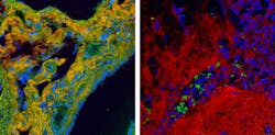
A new form of spontaneous Raman spectroscopy delivers signals 10,000 times stronger than those obtained from spontaneous Raman scattering, and 100 times stronger than signals generated by comparable "coherent Raman" instruments.1 Furthermore, the technique uses a much larger portion of the vibrational spectrum of interest to cell biologists. A version of broadband coherent anti-Stokes Raman scattering (BCARS), the technique is fast and accurate enough to create high-resolution images of biological specimens. Such images contain detailed spatial information on the specific biomolecules present at speeds fast enough to observe changes and movement in living cells.
Most current coherent Raman methods obtain useful signal only in a spectral region containing approximately five peaks with information about carbon-hydrogen and oxygen-hydrogen bonds. The improved method adds to this high-quality signal from the "fingerprint" spectral region, which has approximately 50 peaks—most of the useful molecular ID information.
Also, conventional coherent Raman instruments must tune two separate laser frequencies to excite and read different Raman vibration modes in the sample. The new instrument uses ultrashort laser pulses to simultaneously excite all vibrational modes of interest; this "intrapulse" excitation produces its strongest signals in the fingerprint region. Because too much light will destroy cells, "we've engineered a very efficient way of generating our signal with limited amounts of light. We've been more efficient, but also more efficient where it counts, in the fingerprint region," said chemist Marcus Cicerone, one of the project's researchers from the National Institute of Standards and Technology (NIST; Gaithersburg, MD) working with others from the Cleveland Clinic in Ohio.
Raman hyperspectral images are built up by obtaining spectra, one spatial pixel at a time. The hundred-fold improvement in signal strength makes it possible to collect individual spectral data much faster and at much higher quality than before—a few milliseconds per pixel for a high-quality spectrum vs. tens of milliseconds for a marginal-quality spectrum with other coherent Raman spectroscopies, or even seconds for a spectrum from more conventional spontaneous Raman instruments. Because it's capable of registering many more spectral peaks in the fingerprint region, each pixel carries a wealth of data about the biomolecules present. This translates to high-resolution imaging within a minute or so, whereas, notes NIST electrical engineer Charles Camp, Jr., "It's not uncommon to take 36 hours to get a low-resolution image in spontaneous Raman spectroscopy."
Camp adds, "There are a number of firsts in this paper. Among other things, we show detailed images of collagen and elastin—not normally identified with coherent Raman techniques—and multiple peaks attributed to different bonds and states of nucleotides that show the presence of DNA or RNA."
1. C. H. Camp et al., Nature Photon., 8, 627–634 (2014); doi:10.1038/nphoton.2014.145.

