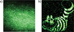
If only light were able to penetrate all the way through the body's tissues, the many advantages of optical imaging could become even more valuable. Now, Spanish researchers have made progress toward that goal with a single-pixel optical system. The scientists, from Jaume I University (UJI; Castellón) and the University of València, used compressive sensing to overcome the fundamental limitations imposed by light scattering in tissue, which severely limits penetration.1 Speaking of standard approaches, Jesús Lancis, a researcher in the Photonics Research Group at UJI, said, "Current knowledge is insufficient for early detection of small lesions located deeper than a millimeter beneath the surface of the mucosa." Lancis is co-author of a paper describing the work.1
The team used a digital micromirror array from a commercial video projector to create a set of microstructured light patterns that are sequentially superimposed onto a sample. They measure the transmitted energy with a photodetector that can sense the presence or absence of light, but has no spatial resolution. Then, they apply a signal processing technique, compressive sensing, to reduce the size of large data files. This allows them to reconstruct the image.
One of the most surprising aspects of the work is the scientists' use of what is essentially a single-pixel sensor. In low-light imaging, it's better to integrate all available light into a single sensor. If the light is split into millions of pixels, each sensor receives a tiny fraction of light, creating noise and destroying the image. "Something similar happens when you try to transmit images through scattering media," he explained. "When we use a conventional digital camera to get an image, we only see the familiar noisy pattern known as 'speckle.' In compressive imaging, since we aren't using pixelated sensors, it should be less sensitive to light scrambling and enable transmission of images through scattering."
Also notable, the team's technique could operate through dynamic scattering. "Most scattering media of interest, like biological tissue, are dynamic in the sense that the scatter centers continuously change their positions with time-meaning that the speckle patterns are 'in motion.' This is ideal for some applications because monitoring the changes of the speckle can reveal information about the sample, but the drawback is that it's a major nuisance to transmit or get images," Lancis pointed out. "Our technique, however, requires no calibration of the medium, and its fluctuations during the sensing stage don't limit imaging ability."
"Our next goal," said Lancis, "is to break the barriers of light penetration depth inside a scattering medium with the state-of-the-art, megapixel-programmable spatial light modulators used in consumer electronics." To do this, the team will need to demonstrate that their technique works, even when the sample is embedded inside the tissue.
1. E. Tajahuerce et al., Opt. Exp., 22, 14, 16945–16955 (2014).

