MULTIPHOTON IMAGING/SPECTROSCOPY: Iterative, nonlinear approach images noninvasively through intact skull

Nov. 13, 2014
A major limitation of optical measurement in biological tissue is that imaging depth is limited by aberration and light scattering. Adaptive optics (AO) and other methods can help to overcome these factors, but impose their own limitations.

A major limitation of optical measurement in biological tissue is that imaging depth is limited by aberration and light scattering. Adaptive optics (AO) and other methods can help to overcome these factors, but impose their own limitations. To meet these challenges, Meng Cui and Lingie Kong of Howard Hughes Medical Institute's Janelia Research Campus have developed an iterative multiphoton adaptive compensation technique (IMPACT), which uses iterative feedback and nonlinearity to quickly measure wavefront in a highly scattering environment.1

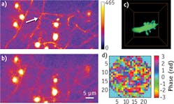
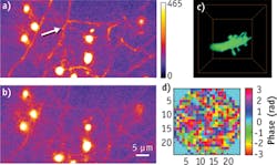
Maximum intensity projection of S1 layer-5 neurons at 654–662 μm depth under the dura of an awake mouse, imaged with correcting both system- and tissue-induced wavefront distortions (a), and system-induced distortion only (b), respectively. The arrow points out a spine, which is invisible in (b). The soma is marked with the asterisk. A 3D rendering of a neuron dendrite was imaged through the intact skull of adult mice in vivo with IMPACT (c). The thickness of the skull is ~120 μm, and the dendrite is 110 μm beneath the bottom of the skull. Without wavefront correction, the structure is buried in background noise and completely invisible. The compensation wavefront used in (c) is shown in (d).

A new paper reports recent progress of applying IMPACT to functional imaging of the cortex to ~660 μm, through the intact skull, in awake mice. In experiments, the technique's high-speed operation (the wavefront can be updated at ~8 kHz and beating frequency is distributed between 2 and 4 kHz) were shown to work well for imaging awake, head-restrained animals, which is challenging for AO. IMPACT compensates wavefront distortion with a large number of spatial modes and does not require taking multiple images; instead, IMPACT maximizes nonlinear (second, third, and higher order) signals, which derives from the laser focus. But laser power during is similar to the actual imaging power, and experiments on GFP- and YFP-expressing samples showed no photobleaching or phototoxicity.

Although the paper mainly discusses imaging applications, the researchers report that IMPACT can also be used for spectroscopy applications to achieve greater signal-to-noise ratio or shorten the measurement time.

1. L. Kong and M. Cui, Opt. Exp., 22, 20, 23786–23794 (2014); doi:10.1364/OE.22.023786.
Sponsored Recommendations

Melles Griot® XPLAN™ CCG Lens Series

March 19, 2024
IDEX Health & Science sets a new standard with our Melles Griot® XPLAN™ CCG Lens Series fluorescence microscope imaging systems. Access superior-quality optics with off-the-shelf...

Spatial Biology

March 19, 2024
Spatial Biology refers to the field that integrates spatial information into biological research, allowing for the study of biological systems in their native spatial context....

Fluorescent Protein Optical Imaging Considerations

March 19, 2024
What factors should you consider when your incorporate fluorescent proteins in an optical imaging application? Learn more.

Custom-Engineered Optical Solutions for Your Application

March 19, 2024
We combine advanced optical design and manufacturing technology, with decades of experience in critical applications, to take you from first designs to ongoing marketplace success...

Voice your opinion!

To join the conversation, and become an exclusive member of Laser Focus World, create an account today!