OPHTHALMOLOGY/ADAPTIVE OPTICS: Adaptive optics shows earliest evidence of diabetic retinopathy

May 19, 2014
A device able to detect the earliest signs yet of diabetic retinopathy leverages adaptive optics and promises important implications for the millions of Americans dealing with the threat of sight loss related to diabetes.

A device able to detect the earliest signs yet of diabetic retinopathy leverages adaptive optics and promises important implications for the millions of Americans dealing with the threat of sight loss related to diabetes.1 Developed at Indiana University (IU; Bloomington, IN) and designed by professor Stephen Burns, associate dean in the School of Optometry, the device uses micro mirrors with moveable segments to reflect light into the eye and to project moving images of the eye's tiny capillaries onto a large screen. Video allows observation of blood cells moving through vessels, while highly magnified retinal images are pieced together with software to create still images.

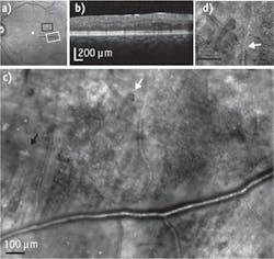
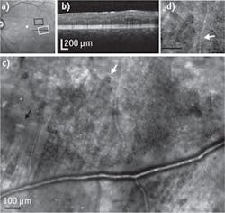
Retinal images from a 31-year-old diabetic subject with moderate nonproliferative diabetic retinopathy: 30 × 30 deg. wide-field scanning laser ophthalmoscopy en face image (a); spectral-domain optical coherence tomography cross-section, with the location as indicated by the black arrow in (a), demonstrating the lack of visible edema or hard exudate (b); a montage of adaptive optics scanning laser ophthalmascopy reflectance images, corresponding to the white rectangle in (a) and showing diffuse capillary remodeling and vessel loops (white arrow), vessel lumen changes, and hard exudates (black arrow) (c); and a blow-up from region of arrow showing a vessel loop as well as changes in small blood vessel wall appearance over space (white arrow) (d). (Image courtesy of Biomedical Optics Express)

"We had not expected to see such striking changes to the retinas at such early stages," says Ann Elsner, a professor and associate dean in the IU School of Optometry and co-author of the study. "We set out to study the early signs in volunteer research subjects whose eyes were not thought to have very advanced disease. There was damage spread widely across the retina, including changes to blood vessels that were not thought to occur until the more advanced disease states."

Because such changes had not been observable in prior studies, it is not known whether improved control of blood sugar or a change in medications might stop or even reverse the damage. Further research can help determine who has the most severe damage and whether the changes can be reversed.

1. S. A. Burns, Biomed. Opt. Exp., 5, 3, 961 (2014).
Sponsored Recommendations

Melles Griot® XPLAN™ CCG Lens Series

March 19, 2024
IDEX Health & Science sets a new standard with our Melles Griot® XPLAN™ CCG Lens Series fluorescence microscope imaging systems. Access superior-quality optics with off-the-shelf...

Spatial Biology

March 19, 2024
Spatial Biology refers to the field that integrates spatial information into biological research, allowing for the study of biological systems in their native spatial context....

Fluorescent Protein Optical Imaging Considerations

March 19, 2024
What factors should you consider when your incorporate fluorescent proteins in an optical imaging application? Learn more.

Custom-Engineered Optical Solutions for Your Application

March 19, 2024
We combine advanced optical design and manufacturing technology, with decades of experience in critical applications, to take you from first designs to ongoing marketplace success...

Voice your opinion!

To join the conversation, and become an exclusive member of Laser Focus World, create an account today!