Research in optical coherence tomography (OCT) technology has recently focused on development of ultrafast (>100 kHz A-line rate) Fourier domain OCT (FD-OCT). While FD-OCT comes in two flavors-spectral domain (SD-OCT), which uses a broadband light source and a high-speed spectrometer, and swept-source (SS-OCT), which uses a high-speed tunable laser and a photodetector for spectral interferogram detection—currently, all FD-OCT systems marketed for clinical ophthalmic use are based on SD.
SD-OCT has several drawbacks, including use of 850 nm light, which is prone to higher tissue scattering than light with longer wavelengths, lower sensitivity, and reduced imaging range (~3.0 mm in air) with rapid sensitivity fall-off (~6 dB/1.5 mm) due to use of a linear sensor array with few pixels. But SS-OCT must clear a number of hurdles before the ophthalmic market will accept it, including receipt of FDA approval. Also, SD-OCT's captured spectral interferogram is highly nonlinear when expressed as a function of optical frequency; achieving longer imaging range and higher axial resolution simultaneously depends on high-speed A/D conversion with stable performance. What's more, phase stability of SS-OCT is relatively poor. And finally, because current commercial ophthalmic systems are based on SD-OCT, the introduction of SS-OCT implies establishment of a new manufacture line.
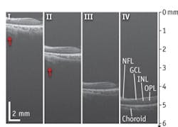
Now, researchers at the University of Washington have developed a high-speed 1050 nm SD-OCT system capable of an axial resolution at ~10 μm in air, an imaging depth of 6.1 mm in air, a system sensitivity fall-off at ~6 dB/3 mm, and an imaging speed of 120,000 A-scans per second.1 Experiments have demonstrated the system's ability to perform phase-resolved imaging of dynamic blood flow within retina, indicating high phase stability, and to image the posterior segment of the human eye with a large field of view (10 × 9 mm2), providing detailed visualization of microstructural features from anterior retina to posterior choroid. The demonstrated system parameters and imaging performances are comparable to those that typical 1 μm SS-OCT would deliver for retinal imaging.
The research team says that implementation of a new prototype high-speed linescan InGaAs camera (Sensors Unlimited Inc.) in the spectrometer is "the critical improvement" of the system, as it provides the necessary boost for imaging depth and system sensitivity, as well as speed to minimize motion artifacts and shorten examination time for a better patient experience.
1. L. An, P. Li, G. Lan, D. Malchow, and R.K. Wang, Biomed. Opt. Exp., 4, 2, 245–259 (2013).

