PHOTOACOUSTIC MICROSCOPY: Isolating signal from photobleached molecules produces super-resolution image

March 18, 2014
Researchers at Washington University in St. Louis (MO) have turned a significant drawback of laser microscopy -- photobleaching -- into an equally significant advantage.

More Biooptics World Articles

INSIDE INSTRUMENTATION: Deep down and label-free: Bioimaging with photoacoustics

OPTICAL COHERENCE TOMOGRAPHY/PHOTOACOUSTIC MICROSCOPY: Single light source correlates OCT and PAM, promising benefits

PHOTOACOUSTIC MICROSCOPY: Isolating signal from photobleached molecules produces super-resolution image

Researchers at Washington University in St. Louis (MO) have turned a significant drawback of laser microscopy—photobleaching—into an equally significant advantage. "Previously when a molecule was prone to bleaching, researchers didn't want to use it because they couldn't get enough information from it," explained postdoctoral researcher Junjie Yao. "Now for us, that is good news."1

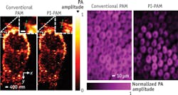
Melanoma cells (left) and rose petal epidermal cells (right) imaged with conventional photoacoustic microscopy (conventional PAM) and photo-imprint PAM (PI-PAM). The resolution is considerably sharper in the PI-PAM images. (Image courtesy of the School of Engineering & Applied Science at Washington University in St. Louis)

The center of a laser beam is so strong that it bleaches a specimen's midpoint, rendering it unable to produce information-packed signal. A second laser pulse probes the molecules in the boundary of the sample, and extracts a relatively weak signal from the bleached area. Yao and colleagues in the lab of photoacoustic microscopy pioneer Lihong V. Wang found that they could collect data from the center of a specimen illuminated by a 200-nm-diameter laser beam by subtracting this boundary area. What's left is a super-resolution image of the 80-nm-diameter center—what they call a photo imprint. The approach effectively shrinks the detection spot, allowing a view of subcellular structure, including features such as mitochondria and cell nuclei.

After each area of the sample is scanned, the researchers create an image. With previous photoacoustic microscopy imaging, the microspheres on the image were blurry. However, with the new photo-imprint photoacoustic microscopy, the result is clear and sharp.

Other scientists working in imaging could apply this method to their own research, Yao said.

1. J. Yao, L. Wang, C. Li, C. Zhang, and L.V. Wang., Phys. Rev. Lett., 112, 014302 (2014).
Sponsored Recommendations

Request a free Micro 3D Printed sample part

April 11, 2024
The best way to understand the part quality we can achieve is by seeing it first-hand. Request a free 3D printed high-precision sample part.

How to Tune Servo Systems: The Basics

April 10, 2024
Learn how to tune a servo system using frequency-based tools to meet system specifications by watching our webinar!

Motion Scan and Data Collection Methods for Electro-Optic System Testing

April 10, 2024
Learn how different scanning patterns and approaches can be used in measuring an electro-optic sensor performance, by reading our whitepaper here!

How Precision Motion Systems are Shaping the Future of Semiconductor Manufacturing

March 28, 2024
This article highlights the pivotal role precision motion systems play in supporting the latest semiconductor manufacturing trends.

Voice your opinion!

To join the conversation, and become an exclusive member of Laser Focus World, create an account today!