ByFlavio Giacobone
In digital microscopy, the importance of the camera cannot be overstated. Recent advances in technology mean the opportunity to select a camera with features that will enhance your particular application.
In digital microscopy, the choice of camera is critical. Since the camera determines the quality of the digitally acquired image, it is key for both analysis and documentation of specimens.
Selecting the most suitable microscope camera very much depends on the desired application. Awareness of the different parameters dictating camera performance, therefore, helps determine the ideal camera for each application, and in turn helps users to reach the full potential of their microscopy system.
Digital camera technology has advanced significantly in recent years and with new technologies regularly emerging, cameras are now available that are suited to individual applications, especially in the life sciences. Let's take a look at four application examples to see how specific camera attributes can contribute to successful outcomes.
True colors
True color reproduction is essential when considering how the variety of hues and intensities enable discernment of different structures in a biological specimen. If color is imposed on the sample by specific staining, for example, it can also lead to diagnostic indications—in which case it is vital to reproduce the exact same color profile on the monitor as one would view through a microscope's eyepieces.
Color cameras measure each pixel hue through a red-green-blue (RGB) filter in front of the sensor. And yet, accurate reproduction has remained a challenge for some time, prior to recent developments in color profiling technologies. Such technologies avoid distracting artifacts such as excessive saturation and color mixing, ensuring faithful reproduction to the extent allowed by the monitor. Some models now even feature native support of the Adobe RGB color space, which allows the camera to interpret a greater range of colors and display them on compatible professional-grade monitors.
On-screen viewing
For imaging specimens, camera speed is critical for enabling the quick location and focusing on regions of interest. It is often the case that the camera will also play a role in displaying an image in real time—for example, when it is necessary to evaluate a sample on-screen, or when presenting and discussing a specimen with an audience.
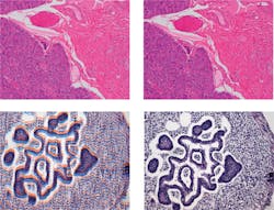
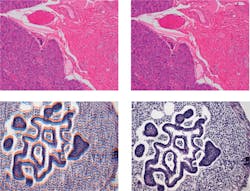
When quality is as important as speed, one feature to look for in a camera is "Full-HD" (high definition), which guarantees the camera is capable of capturing images of at least 2.1 megapixels (Mpixels) at 24 frames per second (frames/s). Too frequently, cameras can exhibit unnatural moving images, and produce problematic striping and color ghosting (see Fig. 1). A remedy for this is "progressive readout" technology, designed to achieve fast and artifact-free images—thus reducing user stress during extended sessions and dramatically improving audience understanding during presentations and discussion sessions.
Switching for sensitivity
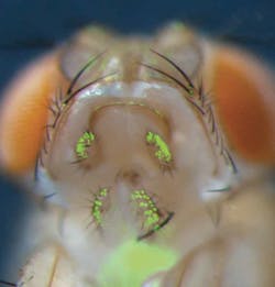
The top priority for low-light imaging and fluorescence applications is sensitivity. Color is not actually required for these techniques, meaning that RGB color filters covering the image sensor chip can be removed to enable superior sensitivity. Furthermore, a dedicated monochrome chip will have a higher sensitivity simply by virtue of having larger pixels, which enable the capture of a greater number of photons and thus produce clear and noise-free images (see Fig. 2). In highly sensitive chips, however, the image noise can be more evident since the measured signal is so low. The most notorious contributor of noise is heat, so it is important to actively cool the camera chip, either with a Peltier-effect plate or with forced air or water flow.
While traditionally it was necessary to choose between color and monochrome options when buying a camera, the arrival of dual-chip cameras has made such decision-making obsolete. The Olympus DP80 camera, for example, houses both dedicated color and monochrome chips, and enables automatic switching depending on the type of snapshot needed. This versatility is incredibly valuable in many labs, and it also presents unique opportunities for overlaying color and fluorescent images with pixel precision.
Documentation and digital archiving
Digital technologies yield many advantages for documentation and archiving, making images highly accessible from a database and, for instance, enabling easy sharing of images—even across the globe-for second opinions. It is important to note that high resolution facilitates the creation of digital sample libraries (see Frontis) since this presents the opportunity for digital zoom, allowing retrospective analysis of archived images (see Fig. 3). Defined both by pixel size (smaller pixels capture finer details) and by the total number of pixels, the camera's resolution is usually summarized in megapixels.
High resolutions are, however, effective only when working at low magnifications. For example, when using a 100x objective, a resolution of just 1 Mpixel is generally sufficient: greater resolution will increase data volume without providing additional information. By contrast, an image captured at 4x magnification might require more than 10 Mpixels to represent all the details from the large field of view. Historically, "pixel shifting" technology allowed the greatest number of pixels to be rendered by a camera, but today, chips with a very high native pixel count are increasingly available.
It is worth noting that the finer pixel size needed for higher resolution has the effect of decreasing a camera's overall sensitivity, but a more versatile solution is "pixel binning," which provides the option to group many small pixels into large "virtual pixels" to increase sensitivity when the application needs it—albeit at the expense of overall resolution.
Core competencies
The camera sits at the very core of ocular-free microscopy and as new advances offer features to enhance a variety of applications, choosing a camera is now a matter of selecting the best options for your projects. For fluorescence imaging, this means taking advantage of the low noise offered by a monochrome chip. For color imaging, it means leveraging color-profiling technologies. For documentation and digital archiving, high resolution is key, while on-screen viewing benefits from progressive readout. Whatever your application, it is wise to choose the latest camera model optimized for that purpose-one that supplies the necessary features to truly enhance the performance of ocular- free microscopy.
In addition, it is important to note that camera capabilities are greatly influenced by software. In fact, it is often possible to enhance image quality by improving the driver software, so make sure that you are using the latest software version available for your camera.
FLAVIO GIACOBONE is a product manager with the Micro-Imaging Solutions Division at Olympus Europa SE & Co. KG (Hamburg, Germany); e-mail: [email protected]; www.olympus-europa.com.


