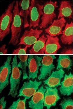
A simple, low-cost method for color-coding individual cells can illuminate 10 times as many biomarkers as is otherwise possible—and thus promises to allow more comprehensive analysis of cell behavior.1 The discovery "opens up exciting opportunities for single-cell analysis and clinical diagnosis," said Xiaohu Gao, associate professor of bioengineering at the University of Washington, and partner in the work with Pavel Zrazhevskiy, a UW postdoctoral associate in bioengineering. The researchers have created a cycle process that allows testing for up to 100 biomarkers in a single cell. Previously, scientists could test for just 10 at a time.
The approach uses quantum dots, and leverages current methods that use a more limited palette to highlight biomarkers for potential disease. It also uses a single tissue sample multiple times, testing for biomarkers in groups of 10 for each round. While such cyclical testing hasn't been explored previously, many quantum dot researchers have worked to tag more biomarkers in a single cell. "Proteins are the building blocks for cell function and cell behavior, but their makeup in a cell is highly complex," Gao said. "You need to look at a number of indicators (biomarkers) to know what's going on." The goal is to highlight hidden patterns and thus show, for instance, why a cell will or won't become cancerous.
To do their testing, the team purchases antibodies that bind with specific biomarkers they want to look for. They match quantum dots with antibodies in solution, which is then injected into a specimen. They then look for fluorescent colors in the cell using microscopy; finding a color means the corresponding biomarker is present.
Following each cycle, they inject a low-pH fluid into the cell tissue to neutralize the color fluorescence, thus "cleaning" the specimen for the next round. Interestingly, the tissue doesn't degrade even after 10 cycles.
For cancer research and treatment, in particular, it's important to be able to look at a single cell at high resolution to examine its details. "When you treat with promising drugs, there are still a few cells that usually don't respond to treatment," said Gao. "They look the same, but you don't have a tool to look at their protein building blocks. This will really help us develop new drugs and treatment approaches."
Gao hopes to find collaborators to work on automating the procedure, and to explore clinical applications, particularly in systems biology, oncology, and pathology.
1. P. Zrazhevskiy and X. Gao, Nat. Comm., 4, 1619 (2013).

