OPTICAL COHERENCE TOMOGRAPHY/AUDIOLOGY: OCT demonstrated for clinical and research application to hearing loss assessment

March 1, 2013
Recently, researchers broke ground on a new application for optical coherence tomography (OCT) when they made the first-ever demonstration of structural and functional imaging of human middle ear micro structures.

Recently, researchers broke ground on a new application for optical coherence tomography (OCT) when they made the first-ever demonstration of structural and functional imaging of human middle ear micro structures. The researchers also demonstrated the diagnostic utility of their approach, and since then at least a couple of groups used the method for both clinical and basic studies.

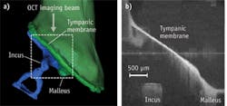
The dotted rectangle (a) shows the middle ear area imaged by OCT (b). (Image courtesy of the Journal of Biomedical Optics)

"Using this method, we could image nanoscale motion of ear drum and ossicles in the human middle ear," explained Hrebesh M. Subhash, lead author on a paper describing the approach.1 This means the approach sheds light on an area no standard hearing test today can measure. Although OCT had been demonstrated previously for imaging the morphology of the human middle ear and tympanic membrane, its application for middle vibrometry in humans had not been reported.

The study demonstrated the use of spectral-domain phase-sensitive (SD-PS) OCT for imaging depth-resolved vibration of middle ear structures through the intact tympanic membrane, and its diagnostic utility on a human cadaver temporal bone. The researchers say this suggests that there may be applications in the diagnosis and preclinical evaluation of middle ear abnormalities. Hearing loss is the world's most common sensory disorder, affecting approximately 17% of American adults.

1. H. M. Subhash et al., J. Biomed. Opt., 17, 6, 060505 (2012); doi:10.1117/1.JBO.17.6.060505.
Sponsored Recommendations

Request a free Micro 3D Printed sample part

April 11, 2024
The best way to understand the part quality we can achieve is by seeing it first-hand. Request a free 3D printed high-precision sample part.

Motion Scan and Data Collection Methods for Electro-Optic System Testing

April 10, 2024
Learn how different scanning patterns and approaches can be used in measuring an electro-optic sensor performance, by reading our whitepaper here!

Case Study: Medical Tube Laser Processing

March 28, 2024
To enhance their cardiovascular stent’s precision, optimize throughput and elevate part quality, a renowned manufacturer of medical products embarked on a mission to fabricate...

Melles Griot® XPLAN™ CCG Lens Series

March 19, 2024
IDEX Health & Science sets a new standard with our Melles Griot® XPLAN™ CCG Lens Series fluorescence microscope imaging systems. Access superior-quality optics with off-the-shelf...

Voice your opinion!

To join the conversation, and become an exclusive member of Laser Focus World, create an account today!