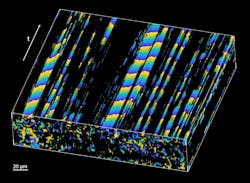
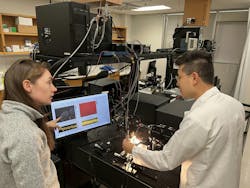
A team of researchers from Baylor College of Medicine (Houston, TX) and Stevens Institute of Technology (Hoboken, NJ) has developed an imaging approach that uses optical coherence tomography (OCT) to observe motile cilia directly in their natural environment (see Fig. 1 and video).
Motile cilia are small hair-like structures covering the epithelial surfaces of multiple organs including airways, lungs, and brain ventricles. Cilia beat in coordination to propel fluids, mucus, and cells along ciliated surfaces.
One of their most important roles lies in the fallopian tubes, says research team leader Dr. Irina V. Larina, a professor of Integrative Physiology at Baylor. There, motile cilia are believed to transport eggs and embryos toward the uterus. “But their specific roles and contributions to infertility and ectopic pregnancy are still unclear,” she says.
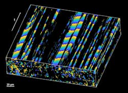
The team’s OCT approach implements spatiotemporal Fourier analysis of intensity fluctuations in images of ciliated samples to first find pixels with significant periodic components corresponding to the cilia locations—the dominant frequency of fluctuations corresponds to the cilia beat frequency. Then, the team maps the phase of the dominant frequency over space and time (see Fig. 2).
A metachronal wave—wavy movements produced by the sequential action of structures like cilia—is created by phase delays in periodic beating between neighboring cilia. This type of phase mapping allows the researchers to visualize and quantify the metachronal wave created by cilia.
Why OCT?
For their study, Larina says OCT was their top-choice starting point because it can best facilitate the study of biological processes in vivo.
“It allows for dynamic volumetric visualization of biological samples with a resolution of a few micrometers (less than the size of one cell) at a depth of 1 to 3 millimeters,” Larina says. “Since it relies on natural tissue contrast and does not require labels, OCT is perfect for in vivo applications.”
Conventionally, high-speed video microscopy is used to expose ciliated surfaces and analyze their dynamics. It’s a more invasive technique that typically involves dissecting an organ of interest and exposing the ciliated surface for imaging. This disrupts the physiological environment of the cilia, resulting in an inaccurate interpretation.
OCT has been part of multiple approaches for motile cilia research and is used for cilia location mapping, cilia beat frequency analysis, and mapping microflows induced by cilia. But the team’s new approach is the first that enables analysis of cilia coordination (metachronal wave) through tissue layers.
“This is important because cilia are working in coordination to move cells and mucus, and proper coordination—not just their beat frequency—is required for the transporting function,” Larina says. “There is currently no other technology that can reach such depth-resolved measurements.”
The research
Ciliary motility is used by microorganisms as well as more complex species including humans; it’s critical for survival. “A complete disruption of ciliary dynamics leads to very early embryonic lethality,” Larina explains. “While subtle abnormalities in ciliary dynamics (motile ciliopathies) lead to serious clinical complications affecting different organ systems.”
Investigation of motile cilia function and regulation “is important for the advancement of healthcare,” Larina says.
Imaging ciliary dynamics is challenging, however, due to motile cilia’s small size. Investigating ciliary beat coordination without directly exposing the ciliated surface was not technologically possible until now.
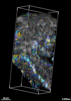
“Our innovation allows us to map cilia metachronal wave propagation within the lumen of a mouse fallopian tube without disrupting the normal physiological environment, which was not previously accessible,” Larina says, and points out that researchers can now map and quantify at micro-scale spatial resolution with a depth-resolved field-of-view (see Fig. 3).
While ciliopathies have been clearly implicated in ectopic pregnancies, the precise role of cilia dynamics in embryo transport remains unknown.
“Our method has the potential to answer the longstanding question about cilia's role in the normal physiology of the fallopian tube and the specific functional consequences of ciliopathies,” Larina says.
In terms of clinical translation in the field of reproduction, fiber-based OCT probes guided through the fallopian tube (falloposcopes) are currently undergoing clinical trials to become standard clinical practice within a few years.
Future implications
The OCT-based approach to imaging motile cilia is poised to also improve the study of physiological processes throughout the body—motile cilia are involved in the regulation of these processes. This platform could be used for various live dynamic investigation of ciliopathies in different organs. Larina says recent advancements in clinical endoscopic OCT imaging in areas including the upper airway make the approach prime for diagnostics of pathologies in the coordination of ciliary activity in the respiratory system.
Another potential research application is related to motile cilia function in the regulation of fluid flows within brain ventricles. Analysis of coordinated ciliary function in the embryonic brain in normal and pathological conditions—for genetic models or in the presence of environmental factors—could provide new insights toward normal brain function and congenital and developmental neurological disorders.
While the team’s new technique is more than promising, Larina says “it will benefit from further optimizations and developments.” She cites, in particular, technological advancements in OCT system design for increased scanning speed, higher resolution, and implementation of multimodality approaches, as well as potential integration with endoscopic imaging.
FURTHER READING
T. Xia, K. Umezu, D. M. Scully, S. Wang, and I. V. Larina, Optica, 10, 1439–1451 (2023); https://doi.org/10.1364/optica.499927.


